


Total:
Expédition: Gratuit
Total (TTC)

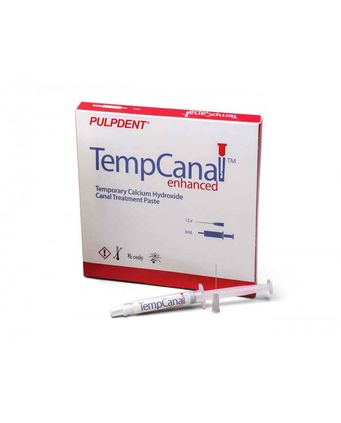
Pâte à l’hydroxyde de calcium pour le traitement canalaire
Caractéristiques :
– Ne sèche pas, durée de travail prolongée
– Formule améliorée Aiguilles 27 ga (type irrigation endo)
– Désinfecte les canaux, protège des inflammations
– pH > 12
Description :
Depuis 1947, avec le développement de la première pâte d’hydroxyde de calcium prémélangée, Pulpdent est devenu le leader en hydroxyde de calcium. Aujourd’hui, avec TempCanal Enhanced, Pulpdent a créé une pâte qui passe à travers les aiguilles endodontiques de 27 gauge permettant ainsi un meilleur contrôle lors du placement dans le canal.
TempCanal Enhanced est plus facile à utiliser, ne sèche pas et sa seringue à bout emoussé empêche les remplissages excessifs. Après une désinfection efficace des canaux, TempCanal Enhanced protège contre les inflammations entre visites et pendant le traitement prolongé des cas compliqués.
Application
Root Canal Therapy
Pulpdent Paste, TempCanal and Multi-Cal are used in endocontics for all the following clinical situations outlined by Dr. G.S. Heithersay:
1. Exudation control; 2. Large periapical lesions; 3. Dressing; 4. Temporary root filling; 5. Apical inflammatory resorption; 6. Inflammatory resorption following trauma; 7. Apical internal resorption; 8. Internal-external root resorption; 9. Perforations; 10. Transverse root fractures; 11. Incompletely developed pulpless teeth
Heithersay GS. Calcium hydroxide in the treatment of pulpless teeth with associated pathology. J Brit Endo Society 1975;8(2):74-93.
Treatment of Abscessed Teeth
Four months after an auto accident in which the patient’s chin hit the steering wheel, the patient presented with loose and painful lower central incisors. We immediately performed root canals and placed TempCanal in the canals to stimulate healing.
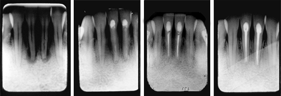
Fig. 1: Radiograph showing abscessed teeth with considerable bone loss.
Fig. 2: Six months after root canals and treatment with TempCanal, radiograph shows bone filling in and healing occurring.
Fig. 3: One year follow up shows healing and obturation with Pulpdent Root Canal Sealer.
Fig. 4: Radiograph taken nine years after final filling shows long term success.
Treatment of Periapical Lesion and Internal Resorption
The patient presented with considerable discomfort and extreme sensitivity to hot, cold and percussion.
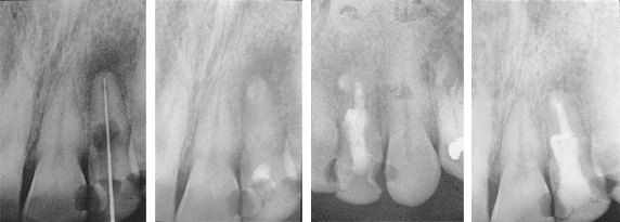
Fig. 1: Diagnostic radiograph shows internal resorption and periapical lesion of the maxillary left lateral. TempCanal was placed at this visit (not shown).
Fig. 2: Radiograph taken 3 months later shows TempCanal in place and periapical healing occurring.
Fig. 3: Radiograph taken after 13 months shows the root canal obturated with Pulpdent Root Canal Sealer using the Pulpdent Pressure Syringe. Note the slight extrusion of sealer beyond the apex and the internal resorption space obturated with sealer.
Fig. 4: Radiograph taken after 19 months shows internal resorption controlled and periapical lesion healed. Note lamina dura. Also shows a portion of the root canal sealer removed and the entire tooth reinforced with Pulpdent HardCord dual cure composite restorative using DenTASTIC as the bonding adhesive.
Treatment of Persistent Periapical Lesion
The Patient had been under treatment for 4 years for a persistent periapical lesion with constant drainage of his left central incisor. Retrograde surgery for removal of the cyst was scheduled. As a temporary measure before surgery, the pulp was removed and TempCanal was placed in the canal. The maxillary left lateral also tested non-vital and was endodontically treated and obturated with Pulpdent Root Canal Sealer at the same visit.
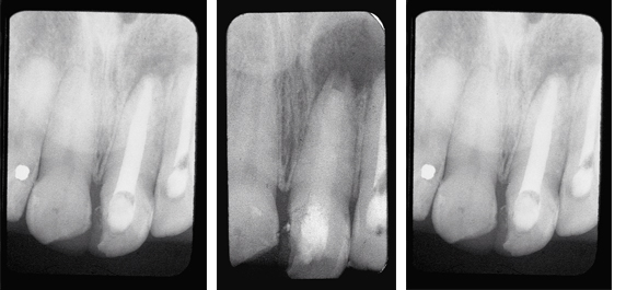
Fig. 1: Radiograph shows periapical lesion involving the maxillary left central and lateral incisors.
Fig. 2: Six weeks following the placement of TempCanal, radiograph shows trabeculation occurring in the periapical area. The surgical procedure was postponed, the TempCanal dressing was changed, and the case was followed until healing of the periapical lesion occurred.
Fig. 3: One year follow up shows healing without surgery and final obturation with Pulpdent Root Canal Sealer using the Pressure Syringe technique.
Hard Tissue Formation
This patient lost his maxillary left central incisor due to a traumatic injury.
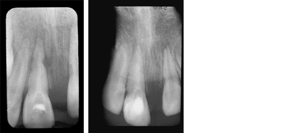
Fig. 1: Radiograph shows external root resorption on the mesial aspect of the maxillary right central incisor. The root canal was filled with TempCanal to promote healing.
Fig. 2: Radiograph taken 3 months later shows remineralization of the mesial aspect of the tooth.
Treatment of Avulsed Tooth
This child presented with avulsed left central and traumatized right central incisors. Two weeks after replantation and splinting, the pulps were removed and Pulpdent Paste was placed in the root canals. The case was followed regularly for 12 months, and the Pulpdent Paste was changed at each visit. After one year, the root canals were obturated with Pulpdent Root Canal Sealer using the Pressure Syringe technique.
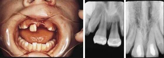
Fig. 1: Photo shows child with avulsed left central and traumatized right central incisor.
Fig. 2: Radiograph taken two weeks following replantation shows replanted tooth, open apices and bone loss. At this visit the root canals were negotiated and Pulpdent Paste was placed as a dressing to stimulate healing and discourage traumatic rejection (not shown).
Fig. 3: Radiograph taken one year after treatment shows Pulpdent Paste in the root canals, apexification and bone fill.


